

Cardiomyopathy
Dilated
Causes
- Myocarditis
- Alcohol
- Prolonged tachycardia
- Pregnancy.
Features
Dilatation and systolic impairment of LV.
Often accompanied by pulmonary hypertension and dilatation of other chambers with MR and TR.
Wall thickness normal.
LV more spherical.
Spontaneous echo contrast and thrombus (slow turbulent flow).
Early closure of AV and delayed closure MV.
As LVEDP rises then will get evidence of diastolic dysfunction.
Hypertrophic
Genetic.
65% asymmetrical (mostly septal but can be apical).
35% symmetrical (need to make sure no other cause like AS or hypertension).
Obstructive or non obstructive.
Systolic function preserved. LV non-dilated with small cavity. EF normal or increased.
Diastolic function impaired.
Obstructive
LVOT increased velocity (significant gradient and turbulent flow - can distinguish AS from HOCM by using PW to see where velocity highest).
Obstructive if gradient >30mmHg (including with exercise).
Sabre shaped LVOT trace (rate of rise in velocity increases throughout systole as the LVOT narrows).
Increased LVOT velocity causes a Venturi effect causing systolic anterior motion (SAM) of the anterior MV leaflet. The leaflet will touch the septum in systole. This may cause a posteriorly directed jet of MR.
Fluttering/early closure of AV.
Velocity increased with valsalva.
Athlete’s heart
Hypertrophied LV (?RV too)
Dilated LV
Normal atrial size
Restrictive
Infiltration or deposition.
Amyloid, haemochromatosis, endocardial fibrosis, sarcoid, glycogen storage disease.
Systolic function preserved. LV non-dilated. (EF preserved but systolic function not normal).
Diastolic function impaired - stiff LV.
LV and RV may be hypertrophied.
Dilated atria and IVC.
Pulmonary hypertension.
Myocardium echo-bright and speckled in amyloid.
Arrhythmogenic RV cardiomyopathy
Dilated RV with dysfunction
Aneurysms of RV free wall.
Echo-bright moderator band and myocardium.
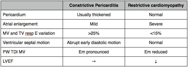
Constrictive Pericarditis vs restrictive cardiomyopathy.