


APICAL VIEWS
A4C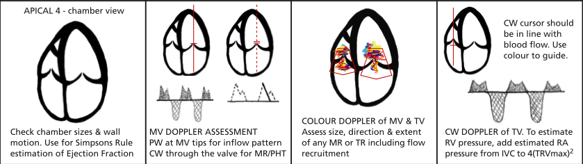
Image from BSE website
Position probe at cardiac apex looking up towards the patients R shoulder and with the marker pointing between the L shoulder and flank. Rotating clockwise from the L shoulder will open up the heart bringing the R side into view.
2D
Overall impression:
- LV and RV fx and RWMAs
- Valve structure and fx
- Dimensions LV, RV, LA, RA
- Wall thickness
- Pericardial effusion
Measure:
Volume of LVd and s, RVd and s, LAs and RAs by Simpsons method (trace around inside chamber and measure length). Ignore papillary muscle.
Basal and mid width RV and length of RV.
M-mode
MAPSE at lateral MV annulus.
TAPSE at lateral TV annulus.
Colour
MV inflow and regurg.
PISA
Vena contracta should be measured in PLAX if possible.
TV inflow and regurg. PISA.
Inter-atrial and ventricular septa.
Doppler
MV
CW
Used to assess MR
- Measure peak velocity of regurg jet (below line)
- Measure rate of ventricular pressure rise (dP/dt). Mark the points where regurg jet velocity reaches 1m/s and 3m/s then measure the time interval between these points
Position sample volume at tips of MV leaflets and measure:
- Peak E and A wave velocity
- E:A ratio
- E wave deceleration time (slope of E decel from peak to baseline)
- Trace round inflow (E and A waves) to measure VTI to calculate mean pressure gradient for MS and regurgitant volume if MR (can do this in CW)
- Measure A duration.
- PV S and D velocity.
- Ar (otherwise termed ‘a’) duration (Ar wave below the line after S and D waves)
- Peak Em, Am, Sm and E/Em ratio. Can substitute ‘prime’ for ‘m’.
CW
TR
- Align probe with any regurgitant jet
- Measure peak velocity of jet
- RVSP - RAP (gradient) = 4 x (Vmax)2
- Gradient + RAP = RSVP
- PASP = RVSP if no PV stenosis
Trace round TV inflow to get the VTI and the mean pressure drop as for MV (can do this in CW)
TDI of lateral TV optional
A5C
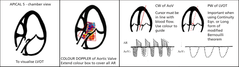
Image from BSE website
Angle the probe slightly anteriorly and the LVOT and aorta will come into view.
2D
Align LVOT and AV with probe
Inspect LVOT (hypertrophy) and AV
Colour
AV - aortic stenosis/regurg
LVOT - flow acceleration (septal hypertrophy)
Septum - VSD
Doppler
CW
Through valve to obtain trace of forward flow +/- regurg
- Measure Vmax from which the gradient can be calculated by the simplified Bernoulli equation. If Vmax LVOT (PW) >1m/s or aorta (CW) <3m/s then need to use full Bernoulli for which you need LVOT VTI from PW as well as aortic VTI from CW.
- Trace round doppler envelope below line to calculate mean pressure gradient (this is the VTI of the aorta). Machine should automatically measure Vmax from a VTI measurement so don’t need to measure Vmax separately.
- If regurg above line, measure pressure half time (downward slope of line).
Position in LVOT where measured LVOT in PLAX.
- Trace around doppler envelope to get VTI for SV and accurate valve area calculations.
- Freeze the trace and measure time between end of aortic outflow and start of mitral inflow - this is the isovolumetric relaxation time (IVRT) used to assess diastolic function. Note BSEs minimum data set says to do this with CW doppler.
A2C
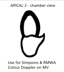 Image from BSE website
From the A4C view rotate the probe anticlockwise so the marker points at the patient’s L shoulder. The R side will disappear leaving a view of only the left side.
2D
Inspect LV dimensions and function
Anterior wall on right of screen; inferior wall on left)
Measure LV area and length (volume) in systole and diastole for EF.
Measure LA area and length.
Colour, CW, PW as for A4C MV
Simpsons for LV volume and EF
A3C / ALAX
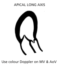 Image from BSE website
Rotate the probe 90 anticlockwise so the marker points at the R shoulder. The LVOT and aorta will come into view.
2D
Anteroseptal wall right of screen; inferolateral left
Don’t do Simpsons in this view
Colour
LVOT and AV
CW and PW for:
- MV
- LVOT
- AV